A Computed Tomography (CT) scan uses X-rays to make pictures of the head and face.
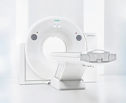
During the test, you will lie on a table that is hooked to the CT scan, which is a large doughnut-shaped machine. Your head will be positioned inside the scanner. The CT scanner sends X-ray pulses through the head. Each pulse lasts less than a second and takes a picture of a thin slice of the head and face. One part of the scanning machine can tilt to take pictures from different positions. The pictures are saved on a computer.
An iodine dye (contrast material) is often used to make structures and organs easier to see on the CT pictures. The dye may be used to check blood flow, find tumors, and look for other problems. Dye can be put in a vein (IV) in your arm. CT pictures may be taken before and after the dye is used.

SOMATOM go.Now